Brain metastases are lesions that appear in the brain when cancer cells from another part of the body travel and implant themselves in nervous tissue. Although brain tumors can be primary or secondary, the clinical approach changes completely when dealing with metastases; the strategy integrates control of the brain tumor and the underlying systemic disease. Early diagnosis and a coordinated treatment plan have a decisive impact on prognosis and quality of life.
Lung, breast, skin (melanoma), kidney, and colon cancers are the most frequent sources of these lesions. If you experience new and persistent headaches, seizures, balance disturbances, weakness on one side of the body, or changes in behavior or memory, it is advisable to seek evaluation by a Neurosurgeon. Timely assessment allows for confirmation of the diagnosis, rapid symptom relief, and determination of whether surgery, radiosurgery, or other therapies are most appropriate for your case.
What Are Brain Metastases and Why Do They Occur?
Brain metastases refer to tumor deposits that originate from a cancer located outside the central nervous system. These cells travel mainly through the bloodstream, overcome biological barriers, and lodge in brain regions with high blood flow, such as the cortico-subcortical junction. Although they may present as single lesions, it is common for multiple foci to appear in different areas of the brain or cerebellum.
When searching for health information, such as brain metastases, this is generally the process being referred to; the primary cancer spreads and establishes secondary growth in the brain parenchyma or leptomeninges. The distinction between a primary tumor and metastasis is key because therapeutic goals, surgical techniques, medications, and follow-up vary according to the origin of the tumor and its molecular biology.
Unlike primary tumors of the nervous system, metastases often present with marked vasogenic edema around the lesion. This edema explains much of the symptoms and, in early stages, may respond well to steroids, providing a window to plan definitive management. Local control (in the brain) must be coordinated with systemic control (in the rest of the body), so teamwork between neurosurgery, medical oncology, and radiotherapy is essential.
Warning Symptoms and How Diagnosis Is Confirmed
Symptoms depend on the size, location, and number of lesions. Some signs require urgent attention, especially if they are of recent onset or progressive.
- Persistent headache, more intense upon waking or different from previous headaches.
- Seizures in an adult with no history of epilepsy.
- Weakness or loss of sensation in part of the body; difficulty speaking or understanding.
- Balance disturbances, double vision, or coordination problems.
- Nausea and vomiting associated with headache or increasing drowsiness.
- Changes in behavior, memory, or personality that interfere with daily activities.
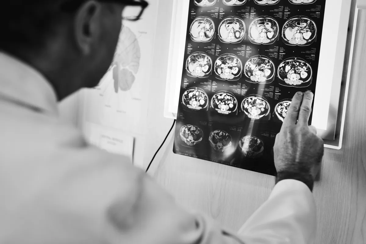
The diagnostic process begins with a detailed neurological evaluation and imaging studies. Contrast-enhanced MRI is the standard for characterizing lesions and planning treatment; CT scans may be useful in emergency settings or when MRI is not available. In selected cases, stereotactic biopsy is required to confirm the histological type, especially if the primary tumor is unknown. In addition, systemic evaluation with CT or PET scans can identify the site of origin and assess the extent.
When secondary brain tumors are suspected, the goal is to confirm the diagnosis precisely, stabilize symptoms (for example, with steroids and antiepileptic drugs when indicated), and prioritize the treatment that offers the best local control with the lowest risk. Individualization is key: not all lesions require surgery, and not all radiotherapy is the same; the choice depends on the size, number of metastases, the patient’s functional status, the type of primary cancer, and response to previous therapies.
Available Treatments in Costa Rica and Key Decisions
Treatment for brain metastases in Costa Rica is defined after multidisciplinary discussion. The goal is to relieve symptoms, preserve neurological function, and achieve the greatest possible tumor control. In our country, high-precision approaches and up-to-date protocols are available to tailor the plan to each patient’s needs.
- Image-guided resective surgery. Indicated for single or dominant, accessible lesions with mass effect. Allows for histological diagnosis and brain decompression to improve symptoms. Modern techniques favor safe resections with neurophysiological monitoring and stereotactic planning. In certain cases, a wide resection followed by radiosurgery to the tumor bed reduces the risk of local recurrence.
- Stereotactic radiosurgery. Useful for small or moderate-sized metastases, single or multiple, and for deep areas or those near eloquent structures. Delivers high doses of radiation with millimetric precision, preserving healthy tissue. Usually performed in one or a few sessions and can be combined with surgery or systemic therapies, depending on tumor biology.
- Whole-brain radiotherapy (WBRT) and cognitive preservation techniques. In selected cases with numerous lesions or meningeal spread, WBRT remains an option. To mitigate neurocognitive effects, hippocampal-sparing techniques and supportive medications are considered. The indication is individualized based on tumor burden and functional status.
- Systemic (oncological) therapies. These include chemotherapy, targeted therapies, and immunotherapy, determined by the type of primary tumor and its molecular alterations. Some molecules achieve adequate penetration into the central nervous system, improving control of metastases. Coordination with medical oncology is crucial to synchronize timing with surgery or radiosurgery.
- Symptomatic management and rehabilitation. Steroids reduce edema and relieve headaches; antiepileptics are indicated if seizures have occurred; support from physical therapy, occupational therapy, and speech therapy helps recover affected functions. Psycho-oncological care and pain management contribute significantly to quality of life.
- Close follow-up and imaging surveillance. After treatment, periodic MRI scans allow for early detection of recurrences and adjustment of the plan. Ongoing communication between teams and with you ensures informed and timely decisions.
Outcomes improve when strategies are combined based on clear clinical and radiological criteria. Factors such as the number of lesions, control of the primary tumor, biological age, and functional scale guide selection. The central goal is for you to maintain your independence and daily activities as long as possible, minimizing side effects.
To receive specialized care and discuss a management plan tailored to your goals and conditions, contact me for a specialized evaluation. A timely consultation clarifies doubts, coordinates priority studies, and accelerates the start of the most appropriate treatment for your particular situation.
